
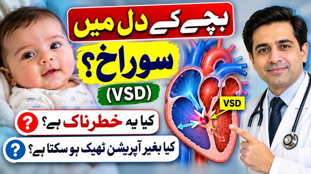
When parents hear that their child has a “hole in the heart,” they naturally become worried. However, not every hole in the heart is dangerous, and many children with this condition can live healthy, active lives.
What Is a Ventricular Septal Defect (VSD)?
A Ventricular Septal Defect (VSD) is one of the most common congenital heart defects in children.
The heart has four chambers: two upper chambers (atria) and two lower chambers (ventricles). Normally, a muscular wall called the ventricular septum separates the two lower chambers.
In children with VSD, there is an opening in this wall that allows blood to pass between the ventricles.
Because pressure is usually higher in the left ventricle, blood flows from the left side to the right side of the heart. This can cause extra blood to travel toward the lungs.
The effect on the child depends mainly on the size and location of the defect and how much extra blood flows through it.
Is VSD a Serious Heart Disease?
The good news is that many small VSDs close naturally as children grow.
VSDs are commonly described as:
1. Small VSD
- Usually causes no significant symptoms.
- The child often feeds and grows normally.
- Many small defects close spontaneously.
- Regular cardiology follow-up may be sufficient.
2. Moderate VSD
- May cause increased blood flow to the lungs.
- Some children experience feeding difficulties or slow weight gain.
- Medicines may be needed to control symptoms.
- Closure may be recommended depending on the child’s condition.
3. Large VSD
- Can cause significant extra blood flow to the lungs.
- May lead to rapid breathing, feeding difficulty, and poor growth.
- Can cause heart failure symptoms in infants.
- Often requires closure during infancy to prevent complications.
Importantly, the size of the hole alone does not determine treatment. Doctors also assess its location, pressure in the lungs, heart chamber enlargement, symptoms, and other associated problems.
What Are the Symptoms of VSD in Babies?
A small VSD may not cause any symptoms. It may be discovered when a doctor hears a heart murmur during a routine examination.
Babies with moderate or large VSDs may develop symptoms such as:
- Fast breathing, especially during feeding.
- Excessive sweating while breastfeeding or bottle-feeding.
- Tiring easily during feeds.
- Taking longer than usual to finish feeding.
- Poor weight gain or failure to thrive.
- Frequent respiratory illnesses.
- Difficulty breathing.
Symptoms of a large VSD often become more noticeable during the first few weeks of life as the resistance in the lung blood vessels naturally falls after birth.
Important: A child with VSD may look completely normal from the outside. Therefore, an echocardiogram is essential when a heart defect is suspected.
How Is VSD Diagnosed?
The most important investigation is echocardiography (heart ultrasound).
An echocardiogram is a safe, painless examination that uses ultrasound waves to visualize the heart.
It helps the pediatric cardiologist determine:
- The location and size of the VSD.
- The direction of blood flow through the opening.
- Whether the heart chambers are enlarged.
- The effect of the VSD on blood flow to the lungs.
- The estimated pressure in the right ventricle and pulmonary circulation.
- Whether other congenital heart abnormalities are present.
An ECG or chest X-ray may occasionally provide additional information, but echocardiography is the main diagnostic test.
Can a VSD Close Naturally?
Yes. Many VSDs, particularly small muscular defects, close on their own.
Some defects close during infancy, while others become smaller over the following years.
However, not every VSD closes naturally.
The chance of spontaneous closure depends on the location, size, and surrounding anatomy of the defect.
Large defects causing significant symptoms should not simply be observed for years in the hope that they will close.
Regular follow-up with a pediatric cardiologist helps determine whether continued observation or treatment is appropriate.
What Are the Treatment Options?
There are three main approaches to managing VSD.
1. Observation and Regular Follow-Up
For small VSDs without symptoms or significant effects on the heart, treatment may not be necessary.
The cardiologist monitors the child’s growth, symptoms, and echocardiographic findings.
2. Medicines
Some babies with larger VSDs develop symptoms of increased blood flow to the lungs or heart failure.
Doctors may prescribe medications, such as diuretics, to reduce symptoms.
Additional nutritional support may also be needed to improve weight gain.
Medicines do not directly close the VSD. They help stabilize the child while the cardiology team determines the appropriate long-term treatment.
3. Closure of VSD
When a VSD causes significant symptoms, heart enlargement, or other complications, closure may be recommended.
There are two main methods.
A. Surgical Closure
Open-heart surgery is the standard treatment for many large or anatomically unsuitable VSDs.
A cardiac surgeon closes the defect, often using a patch.
Modern pediatric cardiac surgery has excellent outcomes in appropriately selected patients.
B. Device Closure (Catheter-Based Treatment)
Certain VSDs can be closed through a catheter procedure without conventional open-heart surgery.
A thin catheter is introduced through a blood vessel, and a specially designed device is positioned across the defect.
However, not all VSDs are suitable for device closure.
Suitability depends on the location of the hole, its relationship to the heart valves and conduction system, the child’s size, and other anatomical factors.
The cardiologist carefully evaluates these details before recommending a procedure.
When Should a VSD Be Closed?
There is no single age that is appropriate for every child.
A large VSD causing significant heart failure or poor growth may need closure during the first few months of life.
Some other defects can be monitored for longer.
The decision depends on the child’s symptoms, growth, echocardiographic findings, and pulmonary blood flow and pressure.
Timely closure of a hemodynamically significant VSD can help prevent long-term complications, including pulmonary vascular disease.
Can a Child With VSD Live a Normal Life?
Yes. Most children with a small uncomplicated VSD or a successfully repaired VSD can enjoy normal growth, education, play, and physical activities.
Many children do not require long-term medication.
Some children need ongoing cardiology follow-up, particularly if there is a residual defect, valve leakage, abnormal heart rhythm, or elevated pulmonary pressure.
Exercise advice should be individualized according to the child’s cardiac condition.
What Should Parents Do at Home?
Parents play an important role in the care of a child with VSD.
- Monitor feeding and weight gain.
- Attend scheduled pediatric cardiology appointments.
- Give prescribed medicines correctly.
- Keep routine childhood vaccinations up to date.
- Maintain good dental hygiene as the child grows.
- Inform the doctor if breathing, feeding, or activity tolerance worsens.
- Avoid giving medicines or restricting physical activity without medical advice.
Most children with VSD do not need antibiotics before routine dental procedures. Antibiotic prevention of infective endocarditis is reserved for specific higher-risk cardiac conditions.
When Should Parents Seek Urgent Medical Attention?
Seek immediate medical assessment if your child develops:
- Severe or rapidly worsening difficulty breathing.
- Blue or grey lips or tongue.
- Inability to feed or unusual exhaustion during feeding.
- Marked lethargy or reduced responsiveness.
- Signs of dehydration, such as significantly fewer wet nappies.
These symptoms require urgent assessment rather than waiting for a routine cardiology appointment.
Frequently Asked Questions
Q1. Is VSD caused by something the mother did during pregnancy?
Usually not. Most VSDs develop during early fetal heart formation without a clearly identifiable cause. Parents should not blame themselves.
Q2. Is VSD hereditary?
Most cases occur without a known family history. However, genetic factors can contribute in some children, and selected families may benefit from genetic counseling.
Q3. Can a small VSD become larger?
Most small VSDs remain stable or become smaller. Changes in blood flow or the appearance of symptoms should be assessed by echocardiography.
Q4. Can children with VSD attend school and play sports?
Most children with small uncomplicated defects or successful repairs can participate in normal activities. Children with significant residual disease require individualized advice.
Q5. Is device closure better than surgery?
Neither method is best for every child. Surgery remains the preferred approach for many VSDs, while catheter-based closure is an option in carefully selected cases.
A Message to Parents
Discovering that your child has a hole in the heart can be frightening. But remember:
Not every hole in the heart needs an operation, and not every child with VSD has a serious heart problem.
The most important step is obtaining an accurate diagnosis and an individualized treatment plan.
With timely assessment, appropriate treatment, and regular follow-up, the outlook for most children with VSD is very good.
This article is intended for general parent education and does not replace an individual medical consultation or emergency assessment.