
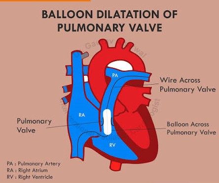
Pulmonary stenosis is a congenital heart condition in which blood flow from the right side of the heart to the lungs is obstructed. In most children, the narrowing occurs at the pulmonary valve—the valve situated between the right ventricle and the pulmonary artery.
The condition may be mild and require only observation, or it may be severe enough to need treatment soon after birth.
How does pulmonary stenosis affect the heart?
The pulmonary valve normally opens freely when the right ventricle pumps blood towards the lungs. In pulmonary valve stenosis, the valve is narrowed, thickened or fused. Consequently, the right ventricle must generate additional pressure to push blood through the restricted opening.
Over time, significant obstruction can cause thickening of the right ventricular muscle and may eventually affect its function. Severe obstruction in a newborn can markedly reduce blood flow to the lungs and require urgent treatment.
What causes pulmonary stenosis?
Pulmonary stenosis usually develops before birth as the baby’s heart forms. In most cases, no single cause can be identified, and it is not caused by anything the parents did during pregnancy.
It may occur:
- As an isolated heart defect
- Alongside other congenital heart abnormalities
- As part of certain genetic syndromes, including Noonan syndrome
- Less commonly, as narrowing below or above the pulmonary valve
What are the symptoms?
The symptoms depend mainly on the severity of the obstruction.
Mild pulmonary stenosis
Most children with mild pulmonary stenosis have no symptoms. The condition may be discovered when a doctor hears a heart murmur during a routine examination.
Moderate or severe pulmonary stenosis
Possible symptoms include:
- Easy tiredness during feeding or physical activity
- Shortness of breath
- Poor weight gain in infants
- Chest discomfort during exertion
- Dizziness or fainting
- Reduced exercise tolerance
- Bluish discolouration of the lips or skin in severe cases
A critically affected newborn may appear blue, breathe rapidly, feed poorly or become unusually sleepy. Such a baby requires immediate medical assessment.
How is pulmonary stenosis diagnosed?
A paediatric cardiologist may use the following tests:
Clinical examination
Pulmonary stenosis often produces a characteristic heart murmur. The child’s oxygen level, pulses and general condition are also assessed.
Echocardiography
An echocardiogram is the main diagnostic test. It is a safe and painless ultrasound examination that shows:
- The structure and movement of the pulmonary valve
- The location of the narrowing
- The pressure difference across the obstruction
- The size and function of the right ventricle
- Any associated congenital heart defects
Electrocardiogram
An ECG records the heart’s electrical activity and may demonstrate strain or thickening of the right ventricle.
Additional imaging or cardiac catheterisation is occasionally required when the anatomy is complex or an intervention is being planned.
Does every child require treatment?
No. Treatment depends on the severity and location of the narrowing, the child’s symptoms, right ventricular pressure and heart function.
Mild stenosis
Children with mild pulmonary stenosis usually do not require medication or a procedure. They need periodic follow-up because the degree of narrowing can occasionally change as the child grows.
Moderate or severe stenosis
Treatment may be recommended when the obstruction is significant, produces symptoms or places excessive pressure on the right ventricle.
Balloon pulmonary valvuloplasty
For suitable pulmonary valve stenosis, balloon valvuloplasty is usually the preferred treatment.
During this catheter-based procedure:
- A thin tube is introduced through a blood vessel, usually in the groin.
- It is guided to the pulmonary valve.
- A special balloon is positioned across the narrowed valve.
- The balloon is briefly inflated to separate the fused valve leaflets and improve blood flow.
The procedure does not require open-heart surgery. Most children recover quickly, although the exact hospital stay depends on their age, condition and response to treatment.
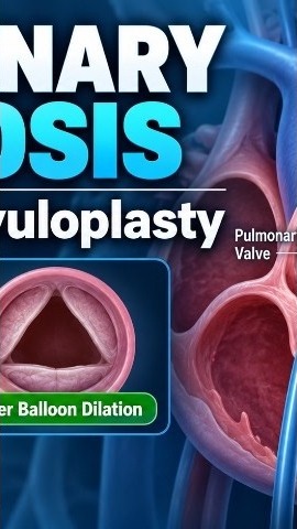
Video: balloon dilation of the pulmonary valve
When is surgery required?
Surgery may be considered when:
- The valve anatomy is unsuitable for balloon treatment
- The valve is markedly thickened or dysplastic
- The obstruction is located below or above the valve
- Other heart defects require surgical correction
- Significant narrowing persists after catheter treatment
The treatment plan is individualized after detailed echocardiographic assessment.
What happens after treatment?
Children require continued follow-up even after successful balloon valvuloplasty or surgery. The cardiologist monitors for:
- Residual or recurrent narrowing
- Leakage through the pulmonary valve
- Right ventricular size and function
- Heart rhythm abnormalities
- Exercise tolerance
Some pulmonary valve leakage is relatively common after balloon treatment. It is often mild, but regular echocardiographic surveillance remains important.
Can a child with pulmonary stenosis live normally?
Most children with appropriately monitored and treated pulmonary stenosis lead active and healthy lives. Children with mild disease may have no meaningful activity limitation.
Exercise recommendations should nevertheless be individualized. A child with severe obstruction, symptoms, abnormal heart function or a recent intervention should obtain clearance from a paediatric cardiologist before participating in competitive sports.
When should parents seek urgent help?
Seek immediate medical care if a child develops:
- Persistent blue lips or skin
- Severe breathing difficulty
- Collapse or fainting
- Marked weakness or unusual sleepiness
- Poor feeding with sweating or rapid breathing
- Severe chest pain during exercise
A reassuring message for parents
Hearing that a child has a narrowed heart valve can be frightening. Fortunately, pulmonary valve stenosis can usually be assessed accurately with echocardiography, and effective catheter-based treatment is available for children who need it.
Early diagnosis, appropriate intervention and regular follow-up provide excellent outcomes for most children.
Medical disclaimer: This article provides general educational information and does not replace an individual medical consultation. The severity and treatment of pulmonary stenosis must be determined by a qualified paediatric cardiologist.
Dr Malik Abid Ali
MBBS, FCPS (Paediatrics), FCPS (Paediatric Cardiology)
Consultant Paediatric and Interventional Cardiologist
Shifa International Hospital, Islamabad