
Introduction: When Your Baby Is Diagnosed with a Heart Condition
Every parent dreams of seeing their child grow up healthy, active, and happy. Hearing that your newborn has a congenital heart defect can be frightening, especially when doctors mention a condition called Tetralogy of Fallot (TOF).
The reassuring news is that Tetralogy of Fallot is a well-recognized and treatable congenital heart condition. With timely diagnosis, appropriate surgery, and lifelong cardiac follow-up, many children with TOF grow up to lead active and fulfilling lives.
In this article, we will explain what TOF is, why some babies appear blue, how the condition is diagnosed, and what treatment options are available.
1. What Is Tetralogy of Fallot (TOF)?
Tetralogy of Fallot is a congenital heart disease, meaning it develops while the baby’s heart is forming during pregnancy.
It accounts for approximately 7–10% of congenital heart defects and is one of the most common causes of cyanotic congenital heart disease.
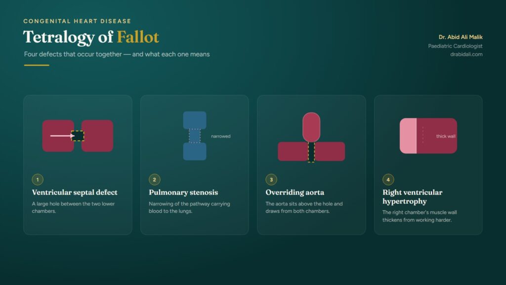
The word tetralogy means a combination of four abnormalities.
The Four Defects of Tetralogy of Fallot
| Heart abnormality | What it means |
|---|---|
| 1. Ventricular Septal Defect (VSD) | A large hole between the two lower chambers of the heart. |
| 2. Pulmonary Stenosis / RVOT Obstruction | Narrowing of the pathway carrying blood from the right ventricle toward the lungs. |
| 3. Overriding Aorta | The aorta is positioned above the ventricular septal defect and receives blood from both ventricles. |
| 4. Right Ventricular Hypertrophy | The muscular wall of the right ventricle becomes thickened because it works against increased resistance. |
Why is this important?
Normally, blood travels from the heart to the lungs to receive oxygen before being pumped to the body.
In TOF, obstruction to blood flow toward the lungs and the presence of a large VSD allow oxygen-poor blood to enter the body’s circulation.
As a result, some babies develop a bluish discoloration of their lips, tongue, or skin, known as cyanosis.
The severity of cyanosis depends mainly on how severely blood flow toward the lungs is obstructed.
2. Why Does Tetralogy of Fallot Occur?
TOF develops during the early stages of pregnancy when the baby’s heart is forming.
In most children, no single identifiable cause is found.
Certain genetic or chromosomal conditions may be associated with TOF, including 22q11.2 deletion syndrome and some other genetic syndromes.
However, many children with TOF have no associated genetic condition.
Important message for parents: In most cases, TOF is not caused by anything the mother or father did during pregnancy.
3. What Are the Symptoms of Tetralogy of Fallot?
Symptoms vary depending on the severity of obstruction to pulmonary blood flow.
Some newborns develop significant cyanosis immediately after birth, while others may initially appear relatively well.
Common symptoms
- Bluish discoloration of the lips, tongue, or skin.
- Rapid breathing, particularly during feeding or crying.
- Difficulty feeding or tiring easily during feeds.
- Poor weight gain.
- Reduced exercise tolerance in older children.
- Episodes of sudden worsening cyanosis, known as Tet spells.
Not every baby with TOF appears blue. Some children with relatively mild obstruction have near-normal oxygen saturation initially. This is sometimes called pink Tetralogy of Fallot.
A normal appearance alone does not exclude TOF.
4. What Is a Tet Spell?
A Tet spell, also called a hypercyanotic spell, is an episode during which the amount of oxygen reaching the body suddenly decreases.
It is particularly important for parents to recognize these episodes because they can become life-threatening.
Warning signs
A baby experiencing a Tet spell may develop:
- Sudden deepening of blue discoloration.
- Rapid or deep breathing.
- Marked irritability or inconsolable crying.
- Unusual weakness or reduced responsiveness.
- Fainting or seizures in severe cases.
These episodes may occur during crying, feeding, waking from sleep, or other periods of increased activity.
What should parents do during a Tet spell?
A suspected Tet spell is a medical emergency.
- Keep the child calm and minimize unnecessary handling.
- Place the infant in a knee-to-chest position, gently bringing the knees toward the chest.
- Seek emergency medical assistance immediately.
- Follow the individualized emergency plan provided by your child’s cardiology team.
For an older child who instinctively squats during an episode, allow them to remain in that position while emergency assistance is arranged.
Do not attempt to treat a severe episode at home or delay emergency care while waiting for symptoms to settle.
5. How Is Tetralogy of Fallot Diagnosed?
The most important diagnostic investigation is echocardiography, commonly called an echo or heart ultrasound.
An echo allows a paediatric cardiologist to examine the structure and function of the baby’s heart.
It helps determine:
- The size and position of the VSD.
- The severity and location of right ventricular outflow tract obstruction.
- The size of the pulmonary valve and pulmonary arteries.
- The relationship of the aorta to the ventricular septum.
- Associated cardiac abnormalities.
- Ventricular function and other features important for surgical planning.
Other investigations may include pulse oximetry, electrocardiography (ECG), chest X-ray, cardiac CT, or cardiac MRI when clinically indicated.
In selected patients, cardiac catheterization may be needed to clarify anatomy or perform an intervention.
Can TOF Be Diagnosed Before Birth?
Yes.
Many cases can be detected during pregnancy through a detailed fetal ultrasound and fetal echocardiography.
Prenatal diagnosis helps the medical team plan delivery at a facility with appropriate neonatal and paediatric cardiac services.
However, some cases are identified only after birth.
6. Can Tetralogy of Fallot Be Treated?
Yes. Tetralogy of Fallot is treatable, and surgical repair is the standard definitive treatment.
Unlike some small ventricular septal defects, classic TOF does not resolve spontaneously.
Treatment aims to improve blood flow to the lungs, close the VSD, and establish a more normal circulation.
A. Initial Medical Management
Newborns with severe obstruction may require specialized treatment soon after birth.
In babies whose pulmonary blood flow depends on the ductus arteriosus, doctors may administer prostaglandin E1 to keep this vessel open temporarily.
Children experiencing hypercyanotic spells require urgent hospital management.
Medical treatment helps stabilize the child but does not replace definitive repair.
B. Complete Surgical Repair
Complete repair generally involves two major steps:
- Closing the VSD
The surgeon closes the hole between the ventricles, usually using a patch.
- Relieving the Obstruction to Pulmonary Blood Flow
The surgeon widens the narrowed pathway between the right ventricle and pulmonary arteries.
Depending on the anatomy, this may involve muscle resection, pulmonary valve repair, or enlargement of the outflow tract.
The surgical approach is individualized to preserve pulmonary valve function whenever feasible.
When Is Surgery Performed?
For many clinically stable infants, complete repair is performed during the first several months of life, commonly around 3–6 months.
However, the timing depends on the baby’s condition and cardiac anatomy.
Newborns with severe cyanosis, duct-dependent pulmonary blood flow, or recurrent Tet spells may require earlier intervention.
Parents should not delay a recommended procedure simply because their baby appears comfortable between episodes.
7. Is Open-Heart Surgery Always Necessary?
Complete surgical repair is required for most children with classic TOF.
However, some babies, particularly those who are premature, very small, or medically unstable, may benefit from an initial procedure that improves pulmonary blood flow before complete repair.
These procedures may include:
| Procedure | Purpose |
|---|---|
| Modified Blalock–Taussig–Thomas shunt | Surgically creates an additional pathway for blood to reach the lungs. |
| Right Ventricular Outflow Tract (RVOT) stenting | Uses a catheter-delivered stent to improve blood flow toward the pulmonary arteries. |
| Ductal stenting | Maintains blood flow through the ductus arteriosus in selected duct-dependent patients. |
These are generally temporary or staged approaches rather than complete correction.
The choice depends on the child’s anatomy, clinical condition, and expertise available at the treating centre.
8. What Is the Success Rate of TOF Surgery?
Outcomes following modern TOF repair have improved considerably.
At experienced paediatric cardiac surgical centres, survival after complete repair is generally above 95% for many patients, although the risk is higher in complex cases and varies between centres.
Factors affecting outcomes include the baby’s age, weight, pulmonary artery development, associated abnormalities, and overall health.
Many children experience a significant improvement in oxygen levels and symptoms after successful repair.
However, TOF repair is not necessarily a once-and-for-all cure.
Lifelong follow-up with a congenital heart specialist remains essential.
9. What Happens After Surgery?
Following complete repair, children usually require monitoring in a specialized cardiac intensive care unit.
The hospital team monitors heart rhythm, oxygenation, ventricular function, fluid balance, and recovery.
Parents receive instructions regarding feeding, medications, wound care, and follow-up before discharge.
The length of hospital stay varies according to the child’s recovery and any complications.
Long-Term Follow-Up
Even after successful repair, some patients may develop:
- Pulmonary valve regurgitation (leakage).
- Residual or recurrent narrowing of the RVOT or pulmonary arteries.
- Right ventricular enlargement or dysfunction.
- Abnormal heart rhythms.
- Residual VSD in a small number of cases.
Some patients require further catheter-based interventions or surgery, including pulmonary valve replacement, later in life.
Regular echocardiography and, when indicated, ECG monitoring, Holter monitoring, cardiac MRI, or exercise testing help identify these problems early.
10. Can a Child with TOF Live a Normal Life?
Many children who undergo successful repair grow up to attend school, participate in family activities, pursue education, and enjoy fulfilling adult lives.
Physical activity recommendations depend on the child’s heart function, oxygen levels, rhythm, and any residual abnormalities.
Children with good surgical results may participate in many forms of physical activity, while those with significant residual disease may require restrictions.
Parents should discuss sports participation with their child’s cardiologist rather than assuming that all exercise is unsafe.
As children grow older, their care should transition to an adult congenital heart disease specialist.
11. Feeding, Growth, and Vaccination
Babies with TOF may require additional nutritional support, particularly before surgery.
Parents should monitor feeding tolerance and weight gain with their healthcare team.
Some infants benefit from smaller, more frequent feeds or individualized feeding plans.
Routine childhood immunizations are generally recommended according to the national schedule, with timing adjusted when clinically necessary around major surgery.
Maintaining good dental hygiene is also important.
Some children with repaired or unrepaired TOF require antibiotic prophylaxis before certain dental procedures because of their risk of infective endocarditis. This depends on their specific cardiac anatomy, repair status, and presence of residual defects.
Your cardiologist can determine whether prophylaxis is indicated.
12. Frequently Asked Questions About TOF
Q1. Is Tetralogy of Fallot a hole in the heart?
TOF includes a large VSD, but it is more complex than an isolated hole because it also involves obstruction to pulmonary blood flow and other structural abnormalities.
Q2. Can TOF close naturally without surgery?
No. Classic TOF does not spontaneously correct itself and usually requires surgical repair.
Q3. Can a baby survive with TOF?
Yes. Many children survive and thrive following timely treatment. However, untreated severe TOF can be life-threatening.
Q4. Is TOF surgery dangerous?
All heart surgery carries risks, but modern surgical techniques have substantially improved outcomes. Individual risk depends on the child’s anatomy and clinical condition.
Q5. Will my child need another operation in the future?
Some children require additional interventions later in life, particularly for pulmonary valve dysfunction or residual obstruction. Others may remain stable for many years without further procedures.
Q6. Can TOF occur in another child in the family?
The recurrence risk is generally low but higher than in the general population. The precise risk depends on whether an underlying genetic condition is present. Genetic counselling may be helpful when planning future pregnancies.
13. When Should Parents Seek Immediate Medical Attention?
Seek emergency care if a child with TOF develops sudden or worsening cyanosis, severe breathing difficulty, fainting, seizures, or reduced responsiveness.
Prompt assessment is also important if the child develops a significant change in feeding, activity, or oxygen saturation compared with their usual condition.
For children who have undergone surgery, new symptoms such as unexplained breathlessness, fainting, palpitations, or reduced exercise tolerance should be assessed by their cardiac team.
A Message to Parents from Dr. Malik Abid Ali
A diagnosis of Tetralogy of Fallot can be overwhelming for any family. However, advances in paediatric cardiology, cardiac imaging, catheter-based interventions, and congenital heart surgery have transformed the outlook for children with this condition.
The most important steps are early diagnosis, timely referral to a specialized paediatric cardiac centre, appropriate treatment, and consistent long-term follow-up.
Every child’s heart is different. Therefore, treatment decisions should be based on a detailed assessment rather than comparisons with other children.
Remember: A child born with TOF can still have a bright future. Early diagnosis, timely treatment, and lifelong cardiac care make a meaningful difference.
About the Author
Dr. Malik Abid Ali
Consultant Paediatric Cardiologist
FCPS Paediatrics | FCPS Paediatric Cardiology
Shifa International Hospital, Islamabad, Pakistan
Dr. Malik Abid Ali specializes in diagnosing and managing congenital and acquired heart diseases in children, with particular interests in neonatal echocardiography, congenital heart defects, and catheter-based paediatric cardiac interventions.
Website: www.drabidali.com
Medical disclaimer: This article provides general educational information and does not replace individualized medical assessment or treatment by a qualified healthcare professional.