
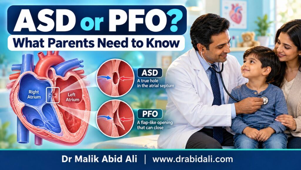
Parents are sometimes told after an echocardiogram that their child has a “hole in the heart.” Two commonly used terms are atrial septal defect (ASD) and patent foramen ovale (PFO).
Although both involve an opening between the heart’s two upper chambers, they are not the same condition. Their importance, follow-up and treatment can be very different.
Understanding the upper chambers of the heart
The heart has two upper chambers:
- The right atrium, which receives oxygen-poor blood from the body
- The left atrium, which receives oxygen-rich blood from the lungs
A wall called the atrial septum separates these chambers.
What is an atrial septal defect?
An atrial septal defect, or ASD, is a congenital heart defect in which part of the tissue separating the two atria has not formed normally. This leaves a true opening in the atrial septum.
Blood usually flows through the ASD from the left atrium to the right atrium. If the opening is significant, extra blood passes through the right side of the heart and into the lungs. Over time, this can enlarge the right atrium and right ventricle and increase blood flow through the pulmonary arteries.
What is a patent foramen ovale?
Before birth, every baby has a natural flap-like opening between the atria called the foramen ovale. It allows blood to bypass the baby’s lungs while oxygen is supplied through the placenta.
After birth, this flap normally closes as pressure inside the heart changes. If it remains partly open, it is called a patent foramen ovale, or PFO.
A PFO is therefore not caused by missing atrial tissue in the same way as an ASD. It is a small flap-like passage left from normal fetal circulation. PFO is common—approximately one in four people may have one—and usually causes no symptoms or health problems.
What is the difference between ASD and PFO?
| Feature | ASD | PFO |
|---|---|---|
| Basic problem | A true deficiency of tissue in the atrial septum | A normal fetal flap that has not completely sealed |
| Blood flow | May allow a significant continuous left-to-right shunt | Usually permits little or no continuous blood flow |
| Effect on the heart | A significant ASD may enlarge the right side of the heart | An isolated PFO usually does not enlarge the heart |
| Follow-up | Depends on the ASD’s size and effect on the heart | Usually no special treatment in a healthy child |
| Closure | May require device closure or surgery | Rarely requires closure during childhood |
Are there different types of ASD?
Yes. The main types include:
Secundum ASD
This is the most common type and is located in the central part of the atrial septum. Depending on its position, surrounding tissue and overall effect on the heart, it may be suitable for closure with a catheter-delivered device.
Primum ASD
This defect is located in the lower part of the atrial septum and may be associated with abnormalities of the atrioventricular valves. It generally requires surgical repair.
Sinus venosus defect
This opening is usually situated near the entry of the upper or lower major vein into the right atrium. It may be associated with abnormal drainage of one or more pulmonary veins and usually requires surgery.
What symptoms can an ASD cause?
Many children with ASD appear completely healthy. The condition may be detected when a doctor hears a heart murmur or when an echocardiogram is performed for another reason.
A moderate or large ASD may sometimes cause:
- Frequent respiratory infections
- Easy tiredness
- Shortness of breath during activity
- Poor weight gain
- Difficulty keeping up with other children
- Excessive sweating or tiring during feeding
- A fast or irregular heartbeat, particularly later in life
Symptoms alone do not determine the importance of an ASD. A child may have few symptoms even when the opening is placing a significant volume load on the right side of the heart.
Does a PFO cause symptoms?
An isolated PFO usually causes no symptoms in babies or children. It normally does not cause poor feeding, recurrent chest infections, breathing difficulty, poor growth or chest pain.
If a child with a PFO has such symptoms, the doctor should look for another explanation rather than assuming that the PFO is responsible.
In uncommon and carefully selected circumstances, a PFO may be considered clinically relevant—for example, when there has been an unexplained blood clot reaching the arterial circulation, decompression illness or another specific medical problem. These situations require specialist assessment and should not be confused with an incidental PFO found on an ordinary echocardiogram.
How are ASD and PFO diagnosed?
Clinical examination
A significant ASD may produce a characteristic heart murmur or splitting of the second heart sound. A PFO usually cannot be detected with a stethoscope.
Echocardiography
An echocardiogram is the main diagnostic test. It is a safe and painless ultrasound examination that can show:
- Whether the opening is an ASD or PFO
- The location and approximate dimensions of an ASD
- The direction and amount of blood flow
- Enlargement of the right atrium or right ventricle
- Pulmonary artery pressure
- Pulmonary venous drainage
- Associated heart abnormalities
- Whether a secundum ASD may be suitable for device closure
Occasionally, a bubble study, transoesophageal echocardiogram or other imaging test is needed to clarify the anatomy.
Can an ASD close naturally?
Some small secundum ASDs diagnosed during infancy become smaller or close spontaneously. Larger defects, primum defects and sinus venosus defects are less likely to close naturally.
The decision to observe or treat an ASD should not be based on a single measurement alone. The cardiologist considers the child’s age, ASD type, blood flow, right-heart enlargement, pulmonary pressure, symptoms and associated abnormalities.
Does every ASD need treatment?
No. A small ASD that does not cause enlargement of the right side of the heart may only require periodic monitoring.
Closure is generally considered when an ASD causes a significant left-to-right shunt or enlargement of the right atrium and right ventricle. Treatment may be recommended even when the child has few obvious symptoms because the aim is to protect the heart and lungs over the long term.
How is an ASD closed?
Device closure through cardiac catheterisation
Many suitable secundum ASDs can be closed without open-heart surgery.
During the procedure:
- A thin tube is introduced through a blood vessel, usually in the groin.
- The catheter is guided into the heart.
- A specially designed closure device is positioned across the ASD.
- The device is released after its position and stability have been carefully checked.
- Heart tissue gradually grows over the device.
The suitability for device closure depends on the ASD’s type, size, position and surrounding rims of tissue. Not every ASD can or should be closed with a device.
Surgical closure
Surgery may be recommended when:
- The ASD is too large or unsuitable for a device
- There is insufficient supporting tissue around the opening
- The defect is a primum or sinus venosus type
- Pulmonary veins drain abnormally
- Another heart abnormality also requires surgical correction
During surgery, the opening is closed directly or with a patch.
Does a PFO need to be closed?
An isolated PFO found incidentally in an otherwise healthy child usually does not require medication, activity restriction or closure. Paediatric evidence for closing a PFO solely for conditions such as migraine remains limited, so such decisions must be individualized.
Parents should not assume that a PFO requires the same treatment as an ASD.
What happens after ASD closure?
Follow-up with a paediatric cardiologist is important after either device or surgical closure. The doctor may monitor:
- Device or patch position
- Any residual blood flow
- Heart chamber size and function
- Heart rhythm
- Pulmonary artery pressure
- Recovery and physical activity
After device closure, medicines such as aspirin may be prescribed for a limited period. The child’s cardiologist will also advise about dental care, infection prevention and when unrestricted activity can be resumed.
Parents should not stop prescribed medication without consulting the treating cardiologist.
Can children with ASD or PFO live normally?
Most children with a small ASD, an isolated PFO or a successfully repaired ASD live active and healthy lives.
Children with an uncomplicated PFO usually need no activity restriction. Exercise advice for a child with ASD should be individualized if there is significant blood flow through the defect, pulmonary hypertension, an abnormal heart rhythm or a recent procedure.
When should parents seek urgent medical care?
Seek urgent medical assessment if a child develops:
- Severe breathing difficulty
- Persistent blue or grey discoloration
- Fainting or collapse
- Sudden weakness or difficulty speaking
- Severe chest pain
- A persistently rapid or irregular heartbeat
- Marked deterioration after a cardiac procedure
These symptoms are not typical of a simple PFO and require proper medical evaluation.
A reassuring message for parents
Finding an opening between the upper chambers of a child’s heart can be worrying, but PFO and ASD are not interchangeable diagnoses.
A PFO is usually a harmless remnant of fetal circulation and commonly needs no treatment. An ASD is a true defect in the atrial septum, but many small ASDs can be observed safely. When a significant secundum ASD requires treatment, it can often be closed through cardiac catheterisation without open-heart surgery.
A detailed echocardiogram and assessment by a paediatric cardiologist can determine the exact diagnosis and the safest plan for your child.
Medical disclaimer: This article provides general educational information and does not replace an individual medical consultation. The significance and treatment of an ASD or PFO must be determined by a qualified paediatric cardiologist.
Dr Malik Abid Ali
MBBS, FCPS (Paediatrics), FCPS (Paediatric Cardiology)
Consultant Paediatric and Interventional Cardiologist
Shifa International Hospital, Islamabad